


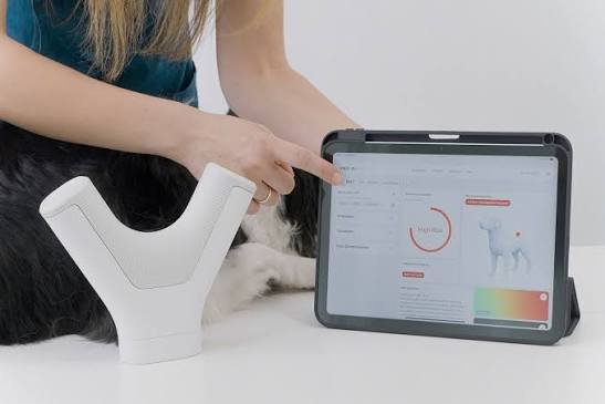
A peer-reviewed clinical evaluation published in Veterinary Sciences demonstrates that HT VET’s VISTA iQ handheld imaging scanner can accurately subclassify specific canine skin masses—specifically differentiating benign lipomas from malignant mast cell tumors (MCTs)—at the point of care. Utilizing Heat Diffusion Imaging (HDI) coupled with advanced artificial intelligence algorithms, the device assesses thermal conductivity signals during a non-invasive, 40-second scan.
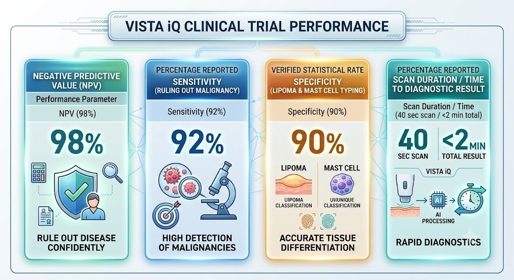
In a multi-center study encompassing over 1,000 subcutaneous and dermal masses across hundreds of canine patients, the platform achieved a 98% Negative Predictive Value (NPV) and 92% sensitivity in ruling out malignancy, while delivering 90% specificity for subclassifying common tumor types. This technology addresses a critical bottleneck in veterinary oncology by enabling rapid, nurse-led triage, eliminating unnecessary invasive procedures for benign fat deposits, and accelerating immediate treatment interventions for aggressive malignancies.
Clinical Challenge: Undiagnosed Dermal & Subcutaneous Masses
Dermal and subcutaneous lumps represent one of the most frequent presentation reasons in general canine practice. Approximately 15% of all superficial masses in dogs are malignant, with mast cell tumors accounting for a major proportion of cutaneous cancers. Conversely, benign lipomas account for a vast majority of non-cancerous subcutaneous swellings.
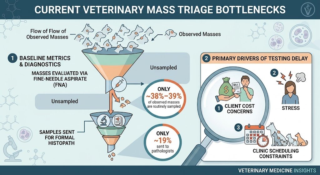
Due to the labor-intensive nature of traditional Fine Needle Aspiration (FNA), client financial pushback, and patient anxiety, veterinary teams frequently resort to a “watchful waiting” approach. This practice risks delaying the diagnosis of mast cell tumors—where early surgical excision prior to metastasis is paramount to long-term survival.
How Heat Diffusion Imaging (HDI) & AI Work
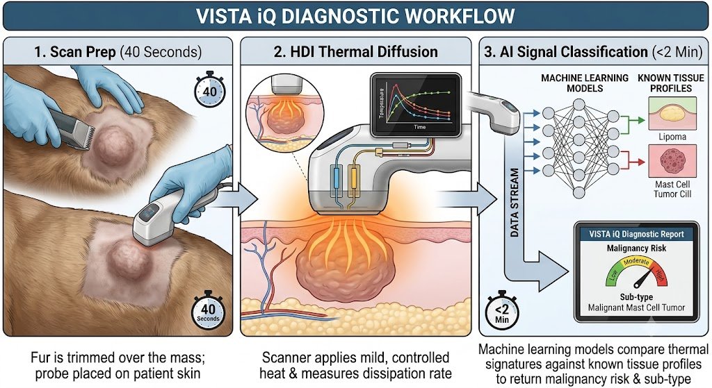
The VISTA iQ device utilizes Heat Diffusion Imaging (HDI), a patented technology that measures the unique thermal transfer properties of living tissue.
-
Thermal Disparity Analysis: Malignant tumors exhibit hyper-vascularization, altered cellular density, and metabolic shifts that dissipate heat at rates drastically different from normal dermis or uniform adipose tissue.
-
Artificial Intelligence Subclassification: The raw thermal diffusion curve is evaluated against a cloud-based algorithm trained on thousands of validated clinical cases. Within two minutes, the system outputs a overall Cancer Risk Assessment (Low, Moderate, High) and, when applicable, an AI-supported Tumor Subclassification identifying lipomas or mast cell tumors.
Study Findings & Diagnostic Performance Metrics
The peer-reviewed study evaluated the device’s diagnostic performance against gold-standard cytology and histopathology.
Key Clinical Outcomes:
-
Confident Rule-Outs: A 98% NPV allows clinicians to confidently reassure owners when a mass receives a “Low Risk” designation, reducing unnecessary invasive biopsies for classic lipomas.
-
Early Mast Cell Interception: High-risk notifications with a mast cell classifier prompt immediate cytology, staging, and surgical planning before the mass enlarges or metastasizes.
Practice Management & Operational Value
Integrating point-of-care AI imaging provides distinct operational and financial benefits for general veterinary practices:
-
Staff Utilization & Nurse-Led Triage: The non-invasive nature of the 40-second scan enables veterinary nurses and technicians to conduct initial screening during routine wellness exams or intake appointments.
-
Client Compliance & Transparency: Owners hesitant to consent to sedation or needle aspirates are far more likely to approve a rapid, painless thermal scan. Clear digital reports build trust and justify immediate follow-up care when high risk is detected.
-
Streamlined Clinical Decisions: By screening every “lump and bump,” practices can significantly increase their diagnostic yield for actionable tumors while improving patient throughput.

