HomeCompanion AnimalsFNA Use in Canine Lumps: Survey Highlights Veterinary Diagnostic Gap

FNA Use in Canine Lumps: Survey Highlights Veterinary Diagnostic Gap

Fine needle aspiration (FNA) is a minimally invasive diagnostic technique that can help veterinarians investigate skin and subcutaneous masses in dogs. Yet survey findings discussed by the American Animal Hospital Association (AAHA) point to a continuing gap between the availability of this diagnostic approach and its routine use in general veterinary practice.

The survey coverage highlighted by AAHA reports that only 29% of companion-animal veterinarians frequently use FNA when initially evaluating canine lumps. The finding raises questions about how consistently suspicious masses are investigated at first presentation and what factors influence diagnostic decisions.

FNA Use in Canine Lumps

Fine Needle Asoirations – Value

Skin and subcutaneous masses are common in dogs. Many are benign, but some are malignant, and appearance or palpation alone cannot reliably distinguish between the two. Delayed investigation can postpone diagnosis and may allow some cancers to progress before treatment begins.

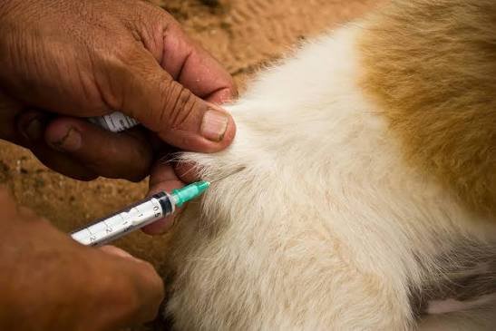
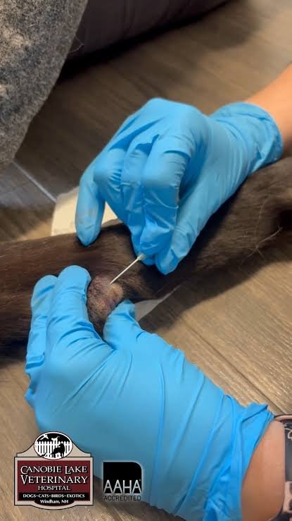
FNA can provide an initial cytological assessment by collecting cells from a mass using a fine needle. The procedure is generally quick and minimally invasive, and samples may be evaluated in-house or submitted to a diagnostic laboratory. It can help guide decisions about monitoring, further testing, biopsy or referral.

However, FNA is not definitive for every mass. Some lesions yield insufficient or poorly representative samples, and cytology may not provide the tissue architecture needed for a definitive diagnosis. Histopathology or additional testing may therefore be required, depending on the lesion and clinical findings.

Why FNAs are Not Effectively Used

The reasons for inconsistent use are multifactorial. Client concerns about cost or discomfort, time constraints, access to cytology services and clinician confidence can all influence whether sampling is recommended or performed. Clear communication about the purpose and limitations of FNA may help owners make informed decisions.

For veterinary practices, establishing a consistent approach to documenting and assessing new masses could improve clinical workflows. This may include recording lesion size and location, discussing sampling options with owners, setting follow-up intervals and identifying cases that warrant more extensive diagnostics.

The industry implications extend to diagnostic laboratories, cytology services, veterinary education and oncology referral networks. Earlier investigation can support more timely clinical decisions, although improved outcomes depend on the type of tumour, diagnostic accuracy and access to appropriate treatment.

The 29% figure should be interpreted in the context of the original survey’s population and definition of “frequent use.” It is not a direct estimate of the proportion of canine lumps that are cancerous or remain undiagnosed.

Animal Health India Editorial Team
Animal Health India Editorial Teamhttps://animalhealthindia.com
Animal Health India (AHI) is an independent news and intelligence platform covering the global animal health, veterinary, livestock, poultry, companion animal and pet food sectors. Our editorial team comprises veterinary journalists, animal health professionals, regulatory affairs specialists and industry analysts with over 30 years of combined experience covering India, Asia, Europe and North America. AHI publishes news, regulatory updates, market intelligence and company news drawn from primary sources including DAHD, EMA, USDA, AVMA and leading veterinary publications worldwide.

RELATED ARTICLES