

A lesion-focused virology study led by Queen’s University Belfast links gallivirus, avian astrovirus and an avian rotavirus to damaged intestinal tissue, opening new avenues for poultry diagnostics and disease-control research.
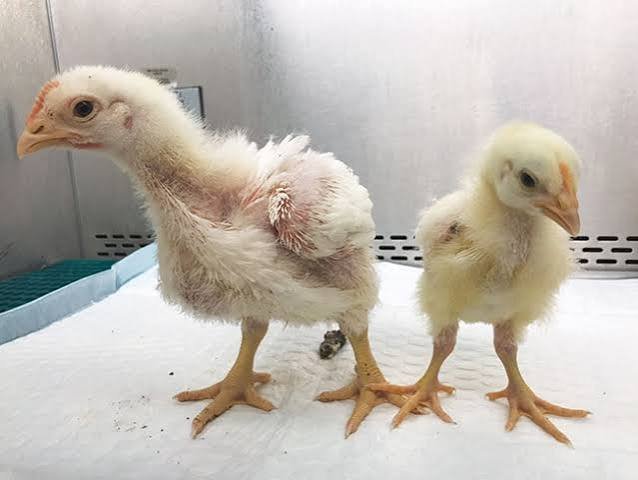
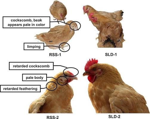
BELFAST, UNITED KINGDOM, 15 October 2026 — Researchers from Queen’s University Belfast and collaborating institutions have identified three candidate enteric viruses associated with intestinal lesions in broiler chickens affected by runting-stunting syndrome (RSS).
The findings, published in the journal Microbiome, could help clarify the infectious factors involved in a disease complex that can undermine growth, flock uniformity and production efficiency.
The study used laser-capture microdissection and viral sequencing to examine tissue directly from affected areas of the gastrointestinal tract. By comparing lesion tissue with non-lesion tissue, researchers were able to investigate which viruses were enriched at sites of damage, rather than relying solely on detection in whole-tissue samples.
The 3 leading candidates were gallivirus, avian astrovirus and a rotavirus. The researchers also confirmed the candidate-virus findings through additional molecular testing and successfully isolated chicken astrovirus in cell culture.
Why runting-stunting syndrome matters to poultry production
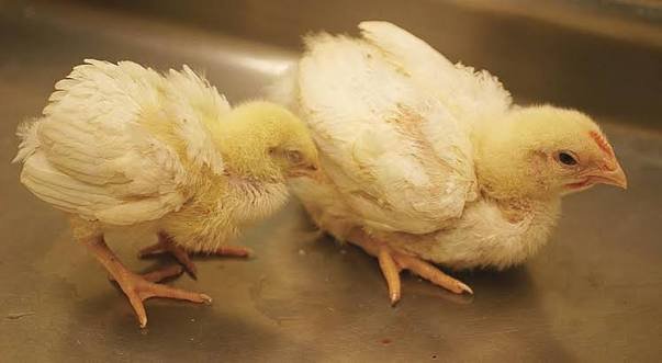
Runting-stunting syndrome is associated with impaired growth and intestinal dysfunction in young chickens. Affected birds may show poor weight gain, uneven body size, abnormal feathering, diarrhoea or the passage of undigested feed. The resulting variation within a flock can complicate management and reduce the efficiency of commercial production.
The economic consequences may include poorer feed utilisation, inconsistent market weights, additional diagnostic and veterinary costs, and greater difficulty achieving uniform processing batches. The scale of the impact varies with the severity and prevalence of disease, production conditions and the effectiveness of management interventions.
The syndrome has been difficult to attribute to a single cause. Previous investigations have detected a range of viruses in affected flocks, but the presence of a virus in a sample does not necessarily mean it is responsible for the observed tissue damage.
This uncertainty has made it challenging to translate pathogen detection into targeted prevention or treatment strategies.